Preimplantation genetic diagnosis
Preimplantation genetic diagnosis (PGD) is a reproductive technology used with an IVF cycle. PGD is used for diagnosis of a genetic disease in early embryos prior to implantation and pregnancy.
Preparation for PGD:
-
Give hormone to the woman to stimulate her ovaries.
-
Remove eggs.
-
Fertilize eggs in the laboratory with sperm.
-
Eggs that are successfully fertilized divide and multiply to form a developing embryo called a blastomere.
-
After three days, the developing embryo will contains about eight cells.
-
Remove one cell for testing.
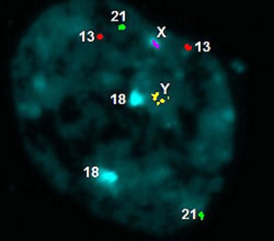
The most commonly applied method for PGD is Fluorescence in situ hybridization (FISH). The embryo cells are fixated on microscope slide and hybridized with DNA probes. Each of these probes are specific for part of a chromosome and are labelled with fluorochrome. The cell is then visualized under the microscope.

Figure 1. Visualization of a cell under the fluorescent microscope (image by Karlovy Vary)